HOW DOES IT WORK? MRI Scanner
FEATURE - 1ST DECEMBER 2016
Dean Booth explains the benefits and complexities of the MRI scanner
Magnetic resonance imaging (MRI) works with the basic elements of the human body by taking advantage of the fact that water molecules in the human body react in a predictable manner when magnetic fields and radio waves are applied to them.
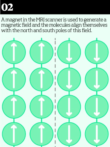
Water molecules within the body have a slight magnetic polarity, with one end being slightly positive and the other slightly negative. For an MRI scan, we first use a superconducting magnet to align the water molecules in relation to the lines of magnetic force.
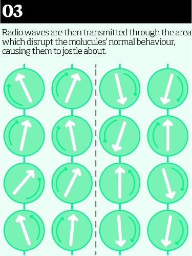
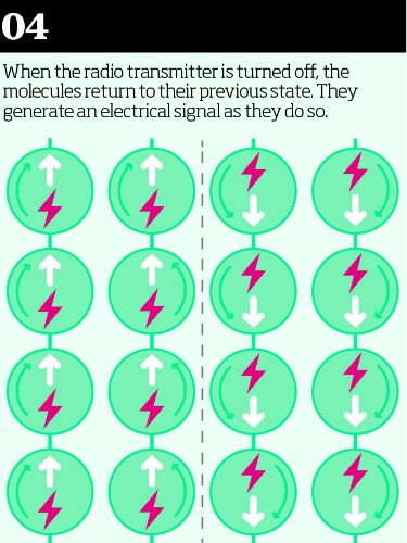
We then transmit radio waves through the area we want to scan. The energy in these waves excites the water molecules, which disrupts their normal behaviour and the levels of energy they possess, causing them to jostle about. The critical part of the process begins after we turn the radio transmitter off. It is while the molecules are relaxing back into their normal state that the scanner’s sensors detect a signal they give off, and it is these signals that are processed by computer to create the image we see. The crucial thing is that we are looking at the change in the energy levels given off by the molecules, not just logging the energy levels themselves.

Different types of tissue, such as fat, muscle or cartilage, will release different amounts of energy in different ways. The scanner records these energy signals throughout the time the molecules are calming down and each reading is given a number—so for example we could have a range of numbers going from 0 to 10. These numbers are then used to create a greyscale image, where black might be represented by 0 and white by 10, and everything in between is a different shade of grey. The more accurate the recording, the more shades of grey you can define and the more detailed your final image will be.
One of the great things about MRI is that while many other scans will give you a static picture of an organ, an MRI can generate a moving image. This really comes into its own with heart scans because a moving image allows you to assess the functionality as well as the anatomy of the heart. The image resolution nowadays is wonderful and we can create a virtual 3D model of what is going on inside the heart, from tiny vessels within the heart right down to the structure of the muscle walls.
One of the things that makes MRI so powerful is that we can change the parameters used to generate the image contrast in order to show us different things and focus in more closely on a particular tissue or area if we need to. The incredible level of spatial resolution you can get on an image allows you to manipulate the image to really narrow in on exactly what you want to see.
MRI can be used to scan most parts of the body. As well as the heart, it is used extensively in brain investigations to look for a wide range of diseases, including dementia. It is also very good at physical structures such as the knee, where we can get detailed images of bone, muscle, cartilage and blood vessels to build a complete picture.
The only real downside of MRI scanning is the time it takes. For example, some brain scans can take over 20 minutes, and the longer the scanner is in use the more expensive it is to do the test. One of the real advances in MRI scanning in recent years has been the speeding up of how long a scan takes. It is better than it was five years ago, but this is always an ongoing process.
As with any medical procedure, there are patient assessments you have to go through before a scan. With MRI, most pacemakers, neurostimulators and ferrous metal implants can be contraindicated. The strong magnetic field can interfere with the function of the implant, and can disrupt, dislodge or heat up components with unfortunate effects. In itself, however, this is a very safe technique. After over 20 years of use all over the world, we are pretty certain that MRI scanning has no short or long term detrimental effects.
Imaging The process of creating visual representations of the interior of a body for clinical analysis
Superconducting magnet An electromagnet made from coils of superconducting wire which is cooled to below -150 °C during operation
Spatial resolution The number of pixels utilised in the construction of a digital image. The higher the spatial resolution, the greater the number of pixels
Ferrous metals Metals that contain iron, and therefore have magnetic qualities
Neurostimulator An implant designed to modulate activity within the nervous system
Contraindication A factor that serves as a reason to withhold a medical treatment, due to the possibility of the patient being harmed
-
HOW TO
HOW TO: Take a pulse
Una Haddick-FLynn, senior nurse at The London Clinic, explains how best to take a pulse.
-
FEATURE
A day in the life: Joana Santos
The daily experiences of a theatre nurse
-
PROFILE
The profile of a pathogen - Campylobacter
Campylobacter is the genus of bacteria known to doctors as one of the most common causes of food poisoning in the world